Background: Neurological disorders known as cerebral palsy affect movement and posture in a large number of patients leading to visual complications. Evaluation of ocular symptoms among children with cerebral palsy leads to timely treatment and care procedures. Objective: This study investigates the prevalence and patterns of ocular disorders among children with cerebral palsy in rural Bangladesh. Methodology: This two-year research period happened at the Centre for the Rehabilitation of the Paralysed from September 2019 to February 2021. Visual acuity tests, cycloplegic refraction and fundoscopic examinations were performed on 170 children who had cerebral palsy. SPSS-26 and MS Excel software were used to conduct the statistical examinations using a p<0.05 level of significance. Results: A high number of refractive errors (75.3%) were detected in the study group and astigmatism (44.7%) turned out to be the most frequently observed condition. Out of all patients examined the researchers discovered strabismus in 34.1% of cases yet exotropia was more common at 18.8% when compared to esotropia at 15.3%. Out of all participants, 10.6% displayed pale optic discs whereas 1.8% exhibited deep cupping during fundoscopic examination. Regular fundus examination results were normal for 87.6% of the total children studied. Conclusion: Cerebral palsy children show high occurrence rates of both refractive errors and strabismus as ocular complications. The evaluation of vision along with prompt interventions by an Ophthalmologist plays an important role in enhancing both the visual performance and life quality of patients. The incorporation of eye care services into rehabilitation treatments and increased patient knowledge about these services will produce better long-term results for children with affected vision.
| Published in | International Journal of Ophthalmology & Visual Science (Volume 10, Issue 2) |
| DOI | 10.11648/j.ijovs.20251002.11 |
| Page(s) | 29-34 |
| Creative Commons |
This is an Open Access article, distributed under the terms of the Creative Commons Attribution 4.0 International License (http://creativecommons.org/licenses/by/4.0/), which permits unrestricted use, distribution and reproduction in any medium or format, provided the original work is properly cited. |
| Copyright |
Copyright © The Author(s), 2025. Published by Science Publishing Group |
Cerebral Palsy, Ocular Manifestations, Refractive Errors, Strabismus, Visual Impairment, Pediatric Ophthalmology, Neuro-ophthalmology
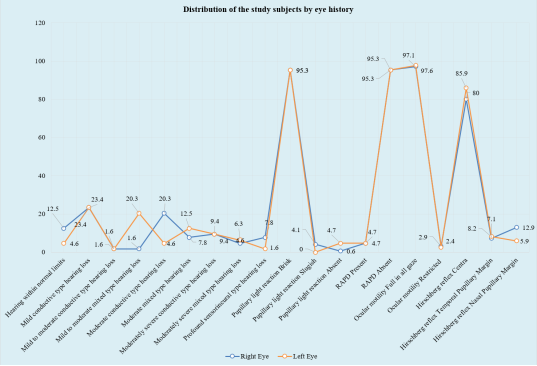
Eye history | Right Eye (n=170) | Left Eye (n=170) | ||
|---|---|---|---|---|
n | % | n | % | |
Pupillary light reaction | ||||
Brisk | 162 | 95.3 | 162 | 95.3 |
Sluggish | 7 | 4.1 | 0 | 0.0 |
Absent | 1 | 0.6 | 8 | 4.7 |
RAPD | ||||
Present | 8 | 4.7 | 8 | 4.7 |
Absent | 162 | 95.3 | 162 | 95.3 |
Ocular motility | ||||
Full in all gaze | 165 | 97.1 | 166 | 97.6 |
Restricted | 5 | 2.9 | 4 | 2.4 |
Hirschberg reflex | ||||
Central | 136 | 80.0 | 146 | 85.9 |
Temporal Pupillary Margin | 12 | 7.1 | 14 | 8.2 |
Nasal Pupillary Margin | 22 | 12.9 | 10 | 5.9 |
Ocular alignment | Number of Subjects | Percentage |
|---|---|---|
Deviation | 58 | 34.1 |
No Deviation | 112 | 65.9 |
Strabismus | Number of Subjects | Percentage |
|---|---|---|
Exotropia | 32 | 18.8 |
Esotropia | 26 | 15.3 |
No Deviation | 112 | 65.9 |
Refractive error | Right Eye (n=170) | Left Eye (n=170) | ||
|---|---|---|---|---|
n | % | n | % | |
Myopia | 8 | 4.7 | 11 | 6.5 |
Hypermetropia | 44 | 25.9 | 44 | 25.9 |
Astigmatism | 76 | 44.7 | 72 | 42.3 |
Fundus | Number of Subjects | Percentage |
|---|---|---|
Normal | 149 | 87.6 |
Deep Cupping | 3 | 1.8 |
Pale disc | 18 | 10.6 |
Ocular morbidity | Number of Subjects | Percentage |
|---|---|---|
Refractive error | 127 | 74.7 |
Strabismus | 58 | 34.1 |
Refractive error and strabismus | 58 | 34.1 |
Pale optic disc | 18 | 10.6 |
Nystagmus | 4 | 2.4 |
Cataract | 2 | 1.2 |
CP | Cerebral Palsy |
VA | Visual Acuity |
RAPD | Relative Afferent Pupillary Defect |
CRP | Centre for the Rehabilitation of the Paralysed |
SPSS | Statistical Package for Social Science |
| [1] | Hallman-Cooper, J. L., & Cabrero, F. R. (2024). Cerebral Palsy. In Treasure Island (FL). StatPearls Publishing. |
| [2] | Jan, M. M. (2006). Cerebral Palsy: Comprehensive Review and Update. Annals of Saudi Medicine, 26(2). |
| [3] | Dilip R Patel, M. N. (2020). Cerebral palsy in children: a clinical overview. Transl Pediatr., 9(Supply 1), S125-S135. |
| [4] | Gulam Khandaker, M. M.-S. (2018). Epidemiology of cerebral palsy in Bangladesh: a population-based surveillance study. Developmental Medicine & Child Neurology, 61(5), 601-609. |
| [5] | Jacy R VerMaas, C. M. (2020). Beyond the eye: Cortical differences in primary visual processing in children with cerebral palsy. Neuroimage Clin., 19(27), 102318. |
| [6] | Elisabeth Mckillop, M. M. (2008). Impairment of vision in children due to damage to the brain: A practical approach. British and Irish Orthoptic Journal, 5, 8-14. |
| [7] | Nikolaos Kozeis, S. J. (2018). Visual Impairment in Cerebral Palsy. In Panteliadis, C. (eds) Cerebral Palsy (pp. 295-302). Springer, Cham. |
| [8] | Saunders, K. J., Little, J.-A., McClelland, J. F., & Jackson, A. J. (2010). Profile of Refractive Errors in Cerebral Palsy: Impact of Severity of Motor Impairment (GMFCS) and CP Subtype on Refractive Outcome. Clinical and Epidemiologic Research, 51, 2885-2890. |
| [9] | Myung Jin Park, Y. J.-M. (2016). Ocular findings in patients with spastic type cerebral palsy. BMC Ophthalmology, 16(195), 1-6. |
| [10] | A Guzzetta, E. M. (2001). Visual disorders in children with brain lesions: 2. Visual impairment associated with cerebral palsy. Eur J Paediatr Neurol., 5(3), 115-9. |
| [11] | Jakob Bie Granild-Jensen, G. R. (2015). Predictors for early diagnosis of cerebral palsy from national registry data. Dev Med Child Neurol., 57(10), 931-5. |
| [12] | A Taylan Ozturk, A. B. (2013). Ocular disorders in children with spastic subtype of cerebral palsy. Int J Ophthalmol., 6(2), 204-210. |
| [13] | Sanjay Marasini, N. P. (2011). Ocular Manifestations in Children with Cerebral Palsy. Optometry & Vision Development, 42(3), 178-182. |
| [14] | Reshma Raj, V. B. (2019). Ocular Evaluation of Patients with Cerebral Palsy. Acta Scientific Ophthalmology, 2(8), 02-07. |
| [15] | S. Shrestha, S. S. (2015). Ocular Morbidity in Children with Cerebral Palsy. Post-Graduate Medical Journal of NAMS, 12(2). Retrieved from |
| [16] | Katoch, S., Devi, A., & Kulkarni, P. (2007). Ocular defects in cerebral palsy. Indian Journal of Ophthalmology, 55(2), 154-156. |
| [17] | Samira Heydarian, M. M.-Y. (2022). Vision Abnormalities in Children and Young Adults With Cerebral Palsy; A Systematic Review. Semin Ophthalmol., 37(4), 471-479. |
APA Style
Bhuiyan, A. R., Sajj, A. B., Sharmin, T. (2025). Ocular Manifestations Associated with Cerebral Palsy Among Rural Bangladeshi Children. International Journal of Ophthalmology & Visual Science, 10(2), 29-34. https://doi.org/10.11648/j.ijovs.20251002.11
ACS Style
Bhuiyan, A. R.; Sajj, A. B.; Sharmin, T. Ocular Manifestations Associated with Cerebral Palsy Among Rural Bangladeshi Children. Int. J. Ophthalmol. Vis. Sci. 2025, 10(2), 29-34. doi: 10.11648/j.ijovs.20251002.11
AMA Style
Bhuiyan AR, Sajj AB, Sharmin T. Ocular Manifestations Associated with Cerebral Palsy Among Rural Bangladeshi Children. Int J Ophthalmol Vis Sci. 2025;10(2):29-34. doi: 10.11648/j.ijovs.20251002.11
@article{10.11648/j.ijovs.20251002.11,
  author = {Ashiqur Rahman Bhuiyan and Abir Bin Sajj and Tohura Sharmin},
  title = {Ocular Manifestations Associated with Cerebral Palsy Among Rural Bangladeshi Children
},
  journal = {International Journal of Ophthalmology & Visual Science},
  volume = {10},
  number = {2},
  pages = {29-34},
  doi = {10.11648/j.ijovs.20251002.11},
  url = {https://doi.org/10.11648/j.ijovs.20251002.11},
  eprint = {https://article.sciencepublishinggroup.com/pdf/10.11648.j.ijovs.20251002.11},
  abstract = {Background: Neurological disorders known as cerebral palsy affect movement and posture in a large number of patients leading to visual complications. Evaluation of ocular symptoms among children with cerebral palsy leads to timely treatment and care procedures. Objective: This study investigates the prevalence and patterns of ocular disorders among children with cerebral palsy in rural Bangladesh. Methodology: This two-year research period happened at the Centre for the Rehabilitation of the Paralysed from September 2019 to February 2021. Visual acuity tests, cycloplegic refraction and fundoscopic examinations were performed on 170 children who had cerebral palsy. SPSS-26 and MS Excel software were used to conduct the statistical examinations using a p<0.05 level of significance. Results: A high number of refractive errors (75.3%) were detected in the study group and astigmatism (44.7%) turned out to be the most frequently observed condition. Out of all patients examined the researchers discovered strabismus in 34.1% of cases yet exotropia was more common at 18.8% when compared to esotropia at 15.3%. Out of all participants, 10.6% displayed pale optic discs whereas 1.8% exhibited deep cupping during fundoscopic examination. Regular fundus examination results were normal for 87.6% of the total children studied. Conclusion: Cerebral palsy children show high occurrence rates of both refractive errors and strabismus as ocular complications. The evaluation of vision along with prompt interventions by an Ophthalmologist plays an important role in enhancing both the visual performance and life quality of patients. The incorporation of eye care services into rehabilitation treatments and increased patient knowledge about these services will produce better long-term results for children with affected vision.
},
 year = {2025}
}
TY  - JOUR
T1  - Ocular Manifestations Associated with Cerebral Palsy Among Rural Bangladeshi Children

AU  - Ashiqur Rahman Bhuiyan
AU  - Abir Bin Sajj
AU  - Tohura Sharmin
Y1  - 2025/04/29
PY  - 2025
N1  - https://doi.org/10.11648/j.ijovs.20251002.11
DO  - 10.11648/j.ijovs.20251002.11
T2  - International Journal of Ophthalmology & Visual Science
JF  - International Journal of Ophthalmology & Visual Science
JO  - International Journal of Ophthalmology & Visual Science
SP  - 29
EP  - 34
PB  - Science Publishing Group
SN  - 2637-3858
UR  - https://doi.org/10.11648/j.ijovs.20251002.11
AB  - Background: Neurological disorders known as cerebral palsy affect movement and posture in a large number of patients leading to visual complications. Evaluation of ocular symptoms among children with cerebral palsy leads to timely treatment and care procedures. Objective: This study investigates the prevalence and patterns of ocular disorders among children with cerebral palsy in rural Bangladesh. Methodology: This two-year research period happened at the Centre for the Rehabilitation of the Paralysed from September 2019 to February 2021. Visual acuity tests, cycloplegic refraction and fundoscopic examinations were performed on 170 children who had cerebral palsy. SPSS-26 and MS Excel software were used to conduct the statistical examinations using a p<0.05 level of significance. Results: A high number of refractive errors (75.3%) were detected in the study group and astigmatism (44.7%) turned out to be the most frequently observed condition. Out of all patients examined the researchers discovered strabismus in 34.1% of cases yet exotropia was more common at 18.8% when compared to esotropia at 15.3%. Out of all participants, 10.6% displayed pale optic discs whereas 1.8% exhibited deep cupping during fundoscopic examination. Regular fundus examination results were normal for 87.6% of the total children studied. Conclusion: Cerebral palsy children show high occurrence rates of both refractive errors and strabismus as ocular complications. The evaluation of vision along with prompt interventions by an Ophthalmologist plays an important role in enhancing both the visual performance and life quality of patients. The incorporation of eye care services into rehabilitation treatments and increased patient knowledge about these services will produce better long-term results for children with affected vision.

VL  - 10
IS  - 2
ER  -
Department of Ophthalmology, Comilla Medical College Hospital, Comilla, Bangladesh
Department of Cornea and Refractive Surgery, Vision Eye Hospital, Dhaka, Bangladesh
Department of Community Medicine, Ad-Din Women’s Medical College, Dhaka, Bangladesh